
Application Note
Cytotoxicity in Cells: Easy Determination Using Lonza ViaLight Plus and ToxiLight BioAssays on the SpectraMax L Microplate Luminometer
By Claire Scholfield Ph.D, Tracy Simmons Ph.D, Susan Gill M.Sc., Lonza Walkerville, Inc.; and Cathy Olsen Ph.D, Molecular Devices.
Introduction
Bioluminescent cytotoxicity assays offer the user increased detection limits, speed, and accuracy, and have been well documented (Crouch et al., 1993). Healthy cells maintain a high constant ATP concentration in culture. During proliferation, the total ATP in the culture increases in line with cell number. Conversely, dying cells are unable to maintain their high ATP level, as synthesis becomes compromised, and so levels are seen to reduce. The level of ATP present within a culture is indicative of the number of viable cells present, and ATP assays have been shown to be one of the most predictive general cytotoxicity methods.
An alternative indicator of cytotoxicity is damage to the plasma membrane. This allows agents to enter the cells (e.g. propidium iodide) and cell components to leak into the surrounding medium. One of the enzymes released when cell membrane damage occurs is adenylate kinase (AK), a ubiquitous protein present in all eukaryotic cells (Squirrell and Murphy, 1997).
The ViaLight® Plus and ToxiLight® Cytotoxicity Assays developed by Lonza exploit these characteristics of cells and enable detection of changes using bioluminescent methods. Both assays have the typical sensitivity of < 10 cells/well. The ViaLight Plus assay has been formulated to produce a long glow signal with a linear decay rate that is irrespective of cell number. The ToxiLight assay is a homogeneous, non-destructive assay enabling the direct assay of wells containing viable and non-viable mixed populations. The assay can also be used on samples of culture media taken from experimental wells, enabling the remaining cultures to be assayed using an alternative method if required. This is a useful feature when samples are precious and multiple analyses are preferred.
Methods
K562 human leukemia cells were serially diluted in complete media and triplicate 100-µL aliquots plated into white-walled luminometer plates. The ATP levels of each well were assayed using the ViaLight Plus assay. Briefly, the cells in each well were lysed by the addition of 50 µL of cell lysis reagent and a 10-minute incubation (at room temperature). Following the lysis, 100 µL of ATP monitoring reagent plus was added to each well and the mixture allowed to equilibrate for 2 minutes. The plate was placed into the SpectraMax® L Luminometer and a 1-second read taken of each well using a preconfigured protocol in the SoftMax® Pro Software (settings shown in Table 1). The results are shown in Figure 1.
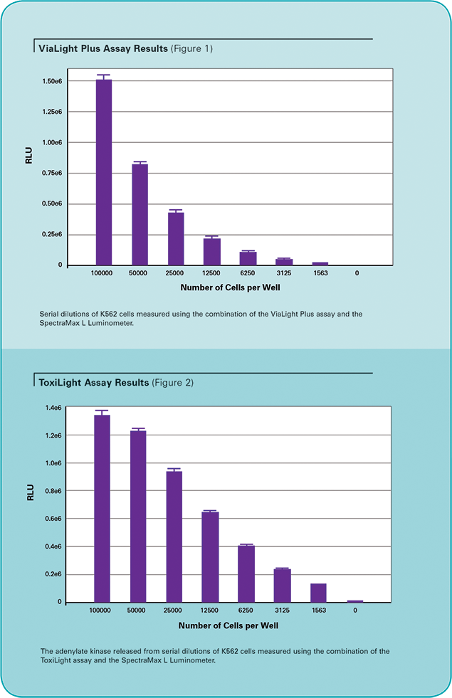
For the ToxiLight assay, 100-µL samples of the serially diluted K562 cells were plated into white-walled luminometer plates. This assay is normally non-destructive to the cells, but for the purpose of demonstrating assay sensitivity, cells were lysed to release their total adenylate kinase (AK). 5 µL of ToxiLight 100% lysis reagent was added to each well, followed by a 10-minute incubation at room temperature. The released AK was assayed using the ToxiLight assay. Briefly, 100 µL of AK detection reagent was added to each well and the mixture incubated for 5 minutes. The plate was placed into the SpectraMax L Luminometer and a 1-second read taken of each well using a preconfigured protocol in SoftMax Pro Software (Table 1). The results are shown in Figure 2.
Both assays provide excellent sensitivity and reproducibility. They are robust, simple to use, and each has its own distinct advantages. The ViaLight assay will show how many viable cells are present by measuring the ATP they release after lysis. The ViaLight assay can show both proliferation and cytotoxicity as compared to an untreated control. As the ToxiLight assay is a non-destructive assay which will only measure AK released from damaged cells, it is a truer indicator of cytotoxicity. These cells, once they are capable of releasing AK, will not recover. This assay will not indicate cells that are healthy or have proliferated.
The ToxiLight assay’s non-destructive sampling protocol enables its use alongside other assays, providing flexibility in experimental design. To demonstrate this, U937 human leukemia cells were treated with camptothecin, a DNA topoisomerase I inhibitor, and were assayed using both ViaLight Plus and ToxiLight reagents as described below.
100-µL samples of U937 cells (5x10⁵ cells/mL) were placed in a 96-well plate, and increasing concentrations of the drug camptothecin were added to the cultures, which were then incubated for 24 hours. Following the incubation step, the samples were assayed using the ToxiLight reagent, following the sampling protocol. 20 µL of cell supernatant was transferred to wells of a white-walled 96-well plate. 100 µL of AK detection reagent was then added to each well, followed by a 5-minute incubation at room temperature. The remaining culture sample was assayed using the ViaLight Plus assay as described above. The assay plates were placed into the SpectraMax L Luminometer and a 1-second read taken for each well using a preconfigured protocol in SoftMax Pro Software (Table 1). The combined results are shown in Figure 3.
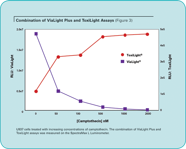
The assumption that fewer viable cells are the result of a cytotoxic event may not always be correct. The combination of the ViaLight and ToxiLight assays is an essential tool to verify this. In this experiment the decreasing RLUs as indicated by the ViaLight Plus assay are accompanied by an increase in the amount of leaked adenylate kinase as measured by the ToxiLight assay. The combination of these assays can provide a more complete picture with minimal extra effort (Table 2). The assays are conducted on the same samples, so the data can be directly compared from well to well and, if required, an extension of the ToxiLight assay’s sampling procedure can provide a useful indicator of the kinetics of cell death.

Conclusions
It is up to users to decide which assay is best suited to their application, but as we have demonstrated here, the combination of the ViaLight Plus and ToxiLight assays provides so much more information that it should be carefully considered. The SpectraMax L Microplate Luminometer and SoftMax Pro Software from Molecular Devices combine well with both the ViaLight Plus and ToxiLight assays from Lonza, providing researchers with a fast and sensitive combination of technologies in the field of cell death. High sensitivity and a large window of detection ensure that cell cytotoxicity is readily detected.
Additional solutions from molecular devices
SoftMax Pro GxP Software provides additional tools and features for users who must demonstrate GLP/GMP compliance with FDA 21 CFR Part 11 requirements. Optional hardware and software validation tools are available to speed and simplify the validation process by making it easier to demonstrate compliance of data collection and analysis.
For increased throughput requirements, the StakMax® Microplate Handling System from Molecular Devices integrates with SpectraMax Microplate Readers and enables automated processing of batches of 20, 40, or 50 microplates.
For more information, please visit www.moleculardevices.com.
References
- . Crouch S, Kozlowski R, Slater K and Fletcher J. (1993). The use of ATP bioluminescence as a measure of cell proliferation and cytotoxicity. J. Immunol. Meth. 160(1) 81–8.
- Squirrel D and Murphy J. (1997). Rapid detection of very low numbers of micro-organisms using adenylate kinase as a cell marker. A Practical Guide to Industrial Uses of ATP Luminescence in Rapid Microbiology P107–113.
By Claire Scholfield Ph.D, Tracy Simmons Ph.D, Susan Gill M.Sc., Lonza Walkerville, Inc.; and Cathy Olsen Ph.D, Molecular Devices.
简介
生物发光细胞毒性检测能提供更高的检 测 限 、 速 度 和 准 确 性 , 已 有 文 献 证 实 (Crouch et al., 1993)。正常细胞在培养过 程中保持高水平的 ATP 浓度。在增殖过程 中,培养物中的 ATP 总量随细胞数量的增 加而增加。相反,死细胞由于合成妥协无 法维持高水平的 ATP,因此认为 ATP 水平 下降。在培养物中 ATP 水平反映了活细胞 数量,ATP 检测已被证实是最重要的预测 一般细胞毒性的方法之一。
另一种细胞毒性指标是细胞膜的损伤。它 允许试剂 ( 如碘化丙钠 ) 进入细胞,细胞成 分会分散到周围的介质中。细胞膜损伤时 会释放腺苷酸激酶 (AK),它是真核细胞中 普遍存在的一种蛋白 (Squirrell and Murphy, 1997)。
龙沙公司开发的 ViaLight Plus 和 ToxiLight 细胞毒性检测试剂盒利用了细胞的这些特 性,通过生物发光的方法检测这些变化。 这两种方法的灵敏度都低于 10 个细胞/孔。 ViaLight Plus 试剂盒能产生一种长时间的 荧光信号,其线性衰减率与细胞数无关。 ToxiLight 试剂盒是一种均相的、对细胞无 损的方法,可以直接测定孔中活细胞和死 细胞的混合总数。该试剂盒也可以用于从 实验板孔中取出的培养基样品,如果需要 还可以用另一种方法对剩余的培养基进行 检测。这对于珍贵样品和需要多种分析的 样品是非常好的选择。
方法
人类白血病细胞 K 562 梯度稀释至培养 基中,三份每份以 100 μL 的倍数加至白 色化学发光板中。采用 ViaLight Plus 试剂 盒检测各孔中的 ATP 水平。每孔中加入 50 μL 细胞裂解液消化细胞,室温孵育 10 分 钟。消化后,每孔加入 100 μL ATP 监测 试剂,混匀后平衡 2 分钟。试剂板放入 SpectraMax L 化学发光微孔板读板机读 板,采用 SoftMax Pro 软件中预设的程序 ( 设置见表 1 ) 每孔读取 1 秒。实验结果见 图 1。
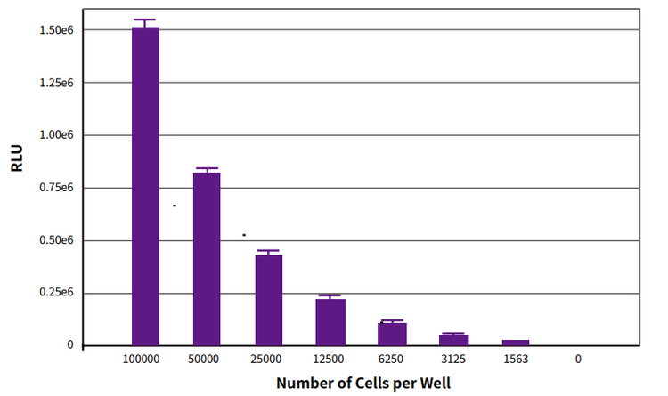
图 1 ViaLight Plus 试剂盒检测结果。梯度稀释 K562 细胞,用 ViaLight Plus 检测 试剂盒和 SpectraMax L 化学发光微孔板读板机读板
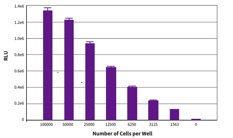
图 2 ToxiLight 检测试剂盒结果。梯度稀释的 K562 细胞释放的 AK 用 ToxiLight 检 测试剂盒和 SpectraMax L 化学发光微孔板读板机检测
采用 ToxiLight 检测试剂盒时,梯度稀释的 100 μL K 562 细胞加入白色化学发光板。 实验本身对细胞无损伤,但是为了达到检 测灵敏度,细胞被裂解释放出总腺苷酸激 酶 (AK)。每孔加入 5 μL ToxiLight 100 % 裂解液,室温孵育 10 分钟。释放出的 AK 用 ToxiLight 检测试剂盒检测。每孔加入 100 μL AK 检测试剂,混匀并孵育 5 分 钟。试剂板放入 SpectraMax L 化学发光 微孔板读板机,采用 SoftMax Pro 软件中 预设的程序 ( 设置见表 1 ) 每孔读取 1 秒。 实验结果见图 2
这两种方法都具有良好的灵敏度和重现 性。它们简单易用,并且各有其独特的优 势。ViaLight 检测试剂盒通过检测消化后 释放的 ATP 来判断有多少活细胞存在。与 未处理的对照组相比,ViaLight 检测试剂 盒可以同时显示细胞增殖和细胞毒性。由 于 ToxiLight 试剂盒是非破坏性实验,它 只能检测从受损细胞释放的 AK,它是一个 更真实的细胞毒性指标。这些细胞一旦释 放出 AK,就无法恢复。所以该试剂盒不能 判断细胞的健康与否或是否有增殖。
ToxiLight 检测试剂盒的非破坏性取样方式 使其能够与其他检测一起进行,实验设计 更加灵活。为了证明这一点,我们使用喜 树碱 ( 一种 DNA 拓扑异构酶 I 抑制剂 ) 处理 U 937 人白血病细胞,并使用 ViaLight Plus 和 ToxiLight 检测试剂盒 ( 如下所述 ) 进行 了检测。
96 孔板中加入 100 μL U 937 细胞 ( 5×10⁵ 细胞/mL ),并往培养基中加入浓度逐渐升 高的喜树碱,培养 24 小时。孵育完成后, 按照试剂盒中取样方法用 ToxiLight 试剂 检测样品。20 μL 细胞上清转入 96 孔白板 中。每孔中加入 100 μL AK 检测试剂,室 温孵育 5 分钟。剩下的培养样品用 ViaLight Plus 检测试剂盒按上面的方法检测。试剂 板放入 SpectraMax L 化学发光微孔板读 板机,采用 SoftMax Pro 软件中预设的程 序 ( 设置见表 1 ) 每孔读取 1 秒。实验结果 见图 3。
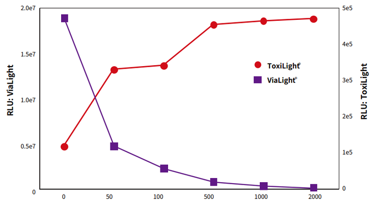
图 3 结合 ViaLight Plus 和 ToxiLight 检测试剂盒结果。U937 细胞用浓度增加的喜 树碱处理。结合 ViaLight Plus 和 ToxiLight 检测试剂盒在 SpectraMax L 化学发光 微孔板读板机上读板
认为活细胞减少是细胞毒性造成的假设并 不总是正确的。同时采用 ViaLight 和 ToxiLight 检测试剂盒检测是验证这一点的 必要工具。在本实验中,ViaLight Plus 检 测试剂盒检测出 RLUs 减少,而 ToxiLight 检测试剂盒检测出腺苷酸激酶释放增加。 结合这两款试剂盒可以以最少的付出得到 更完整的图片 ( 表 2 )。采用相同的样本进 行检测,孔和孔之间的数据可以直接比 较,如果需要,延长 ToxiLight 检测取样 过程能得到有用的细胞死亡动力学指标。
结论
由用户决定哪种检测方法最适合他们的应 用,但是正如本文所示,结合 ViaLight Plus 和 ToxiLight 检测试剂盒检测能够提 供更多的信息,这点是用户需要认真考虑 的。采用 Molecular Devices 的 SpectraMax L 化学发光微孔板读板机和 SoftMax Pro 软件结合龙沙公司 ViaLight Plus 和 ToxiLight 检测试剂盒,为研究人 员提供了一种快速、灵敏的细胞死亡检测 方法。高灵敏度和宽检测窗口,确保细胞 毒性容易检测到。
Molecular Devices 其他解决方 案
SoftMax Pro GxP 能够为用户提供符合 GLP/GMP 合规要求和 FDA 21 CFR Part 11 要求的软件。硬件和软件验证工具能够快 速、简便的完成验证过程,使得验证数据 采集和分析的合规性更加简便。
For more information, please visit www.moleculardevices.com.
参考文献
- . Crouch S, Kozlowski R, Slater K and Fletcher J. (1993). The use of ATP bioluminescence as a measure of cell proliferation and cytotoxicity. J. Immunol. Meth. 160(1) 81–8.
- Squirrel D and Murphy J. (1997). Rapid detection of very low numbers of micro-organisms using adenylate kinase as a cell marker. A Practical Guide to Industrial Uses of ATP Luminescence in Rapid Microbiology P107–113.