
Application Note
Characterization of hERG channel blockers using the FLIPR Potassium Assay Kit on the FLIPR Tetra System
- Functional measurement of K + channel activity in a cell-based assay
- Homogenous no-wash protocol reduces well-towell variation and simplifies the workflow
- Expanded signal window compared to non-homogenous assay
Introduction
Drug-induced inhibition of the human ether-à-go-go-related gene (hERG) ion channel has been related to the susceptibility of patients to potentially fatal ventricular tachyarrhythmia, torsade de pointes. In recent years, a number of FDA-approved drugs were withdrawn from the market due to their off-target effect on hERG. As a result, there has been an increasing need for identifying compounds that block the hERG channel at earlier stages in the drug discovery process. Here we present the utility of a new potassium assay kit on the FLIPR® Tetra High Throughput Cellular Screening System to investigate hERG compound activity. The assay exploits the permeability of thallium (Tl+ ) for potassium (K+ ) channels, which is detected by a novel fluorescence indicator dye. Seven reference hERG blockers are examined in a cell-based assay, and the results are compared to values obtained using the IonWorks Barracuda® Plus Automated Patch Clamp System.
Materials and methods
The FLIPR® Potassium Assay Kit (Figure 1) contains a thallium-sensitive indicator dye. During the initial dye-loading step, the Tl+ indicator dye as an acetoxymethyl (AM) ester enters the cells through passive diffusion across the cell membrane. Cytoplasm esterases cleave the AM ester and relieve its active fluorogenic form. In addition, a proprietary masking dye is applied extracellularly to reduce background fluorescence. To activate the potassium channel, the cells are stimulated with either a mixture of K+ and Tl+ or a ligand in the presence of Tl+ . The increase in fluorescent signal represents the influx of Tl+ into the cell specifically through the potassium channel, and therefore represents a functional measurement of the potassium channel activity. The FLIPR Potassium Assay Explorer Kit (Molecular Devices cat. #R8222) contains Tl+ -sensitive dye, masking dye for homogenous operation, 200 mM K2SO4, 50 mM Tl2SO4, 5X chloride-free buffer, and HBSS + 20 mM HEPES buffer. The kit supports ten 96-, 384-, or 1536-well plates. The assay workflow is shown in Figure 2.
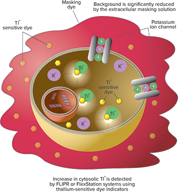
Figure 1. FLIPR Potassium Assay Kit principle.
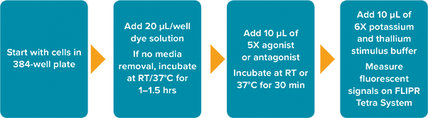
Figure 2: FLIPR Potassium Assay Kit workflow on the FLIPR Tetra System.
Compound preparation
The hydrophobic property of compounds impacts the apparent potency values, likely through non-specific binding to labware. In this study, compounds were first diluted in 100% DMSO, followed by transfer with mixing to HBSS + 20 mM HEPES buffer in glass-lined polypropylene plates immediately prior to assay.
Experimental procedure
Chinese hamster ovary (CHO) cells stably transfected with human Kv11.1 (hERG) ion channel were provided by ChanTest Corporation (Cleveland, OH). Cells are plated at 6,500/well in a 384-well, blackwalled, clear bottom plate two days before the assay in growth medium including selection antibiotics and incubated at 37°C and 5% CO2. Twenty four hours prior to the assay, the growth media is exchanged for induction media containing tetracycline. Four hours prior to the assay, the cells are switched from 37°C to 28°C for enhanced membrane expression of hERG channel. Plates are incubated with dye for one hour at room temperature in the dark.
For pharmacology analysis, hERG channel blocking compounds are added first and incubated for 30 minutes at room temperature. Previously optimized stimulus buffer is added to the well during detection on the FLIPR Tetra System. The 470-495 nm excitation LEDs and 515-575 nm emission filter are used). For comparison, parallel experiments were performed using the FluxOR Assay Kit (Life Technologies). The manufacturer assay protocol was followed when using this kit. Data is acquired at one second intervals for approximately 140 seconds. Data files are exported to GraphPad Prism for analysis.
Assay development is first carried out by determining optimal concentrations of thallium and potassium necessary for stimulation of the hERG channel. Plates are set up to test stimulating buffers containing various concentrations of K2SO4 and Tl2SO4.The Tl+ and K+ buffers are diluted in 1X chloride-free buffer. Since thallium sulfate and potassium sulfate have two equivalents of cation per mole, they are considered as having 2X their respective cation concentrations. The stimulus buffers are added to cells during detection on the FLIPR Tetra System. Signal traces are compared among the concentration combinations to determine the optimal concentrations that provide the largest signal. Here, the optimal (final) Tl+ concentration that provided the largest signal for hERG is 1 mM and the optimal (final) K+ concentration is 10 mM (Figure 3).

Figure 3: Optimization of hERG channel stimulant. Cells were incubated with dye and the stimulant buffers were added during detection on the FLIPR Tetra System. The concentration-dependent response of signal was characterized under different conditions. Optimal signal was obtained from the combination of 1 mM Tl+ and 10 mM K+ (final concentration) stimulant buffer diluted in chloride-free buffer.
Electrophysiology
For comparison purposes, IC50 values for the same set of hERG channel blockers were collected using the IonWorks Barracuda System1 (Figure 4). To capture frequency-dependent compound effects, the voltage protocol is applied five times at 0.1 Hz before and after compound addition. Peak amplitude of hERG tail current at the fifth sweep is used to measure compound effects.

Figure 4. Electrophysiology assay on the IonWorks Barracuda System. Compounds were added at 3X final assay concentration in 1% DMSO and mixed with buffer in the well to achieve a 1X final compound concentration in 0.33% DMSO. Compounds were incubated for five minutes, after which a voltage protocol was applied with a one-second stimulation at +40 mV, followed by a one-second step to -50 mV for measurement of peak tail current.
Results
Seven known hERG blockers were evaluated using the FLIPR Potassium Assay Kit. The concentration response curves are shown in Figure 5. IC50 values compared to data obtained on the IonWorks Barracuda System are shown in Table 1. The rank order of compound potency was preserved between the two assays and IC50 values were within one-half log with good correlation.

Figure 5. Concentration-dependent inhibition of hERG channel by reference compounds.
IC
50
(nM) FLIPR Tetra System
IC
50
(nM) IonWorks Barracuda System
15.3
2
Table 1. Comparison of IC50 values of the FLIPR Potassium Assay Kit on the FLIPR Tetra System and electrophysiology data on the IonWorks Barracuda System.
In addition, three compounds were used to evaluate performance against another thallium-based assay kit, the FluxOR Potassium Ion Channel Assay (Figure 6). The IC50 values are similar between assays as shown in Table 2. However, the signal window provided by the FLIPR Potassium Assay Kit is 2.25 (determined as ratio of response vs. baseline), which is significantly higher than the value for FluxOR (0.5). The negative control is 4 µM terfenadine which blocks the hERG channel response, and the positive control is buffer which elicits maximum response by the hERG channel to the stimulus buffer. Lower well-to-well variability and a larger signal window combine to provide larger Z’ factors as shown in Figure 7. This is likely due to the FLIPR Potassium Assay Kit being a truly homogeneous assay that does not require wash steps or media replacements and therefore shows less well-to-well variability.

Figure 6. Comparison of FLIPR Potassium Assay Kit results to a competitor kit.
IC
50
(nM) FLIPR Potassium Assay Kit
IC
50
(nM) FluxOR Kit
Table 2. Comparison of IC50 values between FLIPR Potassium Assay Kit to a competitor kit.

Figure 7. Comparison of signal dynamic range between the FLIPR Potassium Assay Kit and a competitor kit. Negative control is 4 µM terfenadine; positive control is buffer. The Z’ factor for the FLIPR Potassium Assay Kit = 0.85 and n = 32 compared to the competitor Z’ factor = 0.64 and n = 30.
Conclusion
The FLIPR Potassium Assay Kit measures the functional activity of potassium channels using a homogeneous, no-wash protocol. In this study, we used a set of reference hERG blockers to demonstrate the tight correlation of results from the FLIPR Tetra System with data collected using electrophysiology methods.
In a separate set of experiments, the FLIPR Potassium Assay Kit displayed a significantly larger assay window and a higher Z’ factor when compared to a competitor product. The improved assay quality, in combination with the high-throughput capability of the FLIPR Tetra System, provides a powerful platform for analyzing hERG liability during earlier phases of the drug discovery process.
Compatible with these Molecular Devices systems

References
- Karen Cook, James L. Costantin, and Xin Jiang, Validation of the IonWorks Barracuda System for hERG Ion Channel Assay, Application Note, 2011.
- D. Rampe, et al, A mechanism for the proarrhythmic effects of cisapride (Propulsid): high affinity blockade of the human cardiac potassium channel HERG, FEBS Letters 1997; 417(1): 28-32.
Learn more >>
简介
药物诱导的人类 hERG 离子通道阻滞与患者潜在的致死性室 性快速心律失常的易感性有关。近年来,FDA 批准的一些药物 由于对 hERG 的脱靶作用而退出市场。因此,在药物发现过 程的早期阶段,迫切需要鉴定能够阻断 hERG 通道的化合物。 在此,我们展示在 FlexStation 3 多功能酶标仪上,一种新的 钾离子通道检测试剂盒研究 hERG 化合物活性的用途。这种 分析方法利用了铊离子 (Tl+ ) 的渗透性,通过钾离子通道进入 胞质,然后被一种新型荧光指示剂检测到。7 种参照的 hERG 阻滞剂在中等 通 量 细胞试 验中进 行检 测,并将 FLIPR Tetra System 和 IonWorks Barracuda Plus Automated Patch Clamp System 收集的数据值进行比较。
材料和方法
FLIPR 钾离子通道检测试剂盒 ( 图 1 ) 含有对铊敏感的指示 剂。在起始的染料加样步骤,铊离子指示剂以乙氧基甲基酯 (AM) 的形式通过被动扩散穿过细胞膜进入细胞内。细胞质的 酯酶可切割 AM 酯并释放其活性荧光构型。此外,一种具有 专利的掩蔽染料应用于细胞外以降低背景荧光。细胞被钾离 子和铊离子的混合物或铊离子存在下的配体刺激,可以激活 钾离子通道。荧光信号的增加表示铊离子流入细胞内,确切 地说是通过钾通道流入,因此显示了钾离子通道功能性的活 性 测 量。FLIPR Potassium Assay Explorer Kit (Molecular Devices Cat# R8222) 试剂盒包含铊离子敏感的染料,掩蔽染 料用于均质化操作,200 mM 硫酸钾,50mM 硫酸铊,5x 无氯 缓冲液,HBSS + 20 mM HEPES 缓冲液。此试剂盒支持 10 块 96 孔或 384 孔板。实验流程如图 2。
图 1 FLIPR 钾离子通道检测试剂盒的原理。
图 2 在 FlexStation 3 酶标仪上,FLIPR 钾离子通道检测试剂盒的工作流程。
制备化合物
化合物的疏水性质影响表观效力值,可能是由于与实验器具 的非特异性结合。在这项研究中,化合物首先在 100% DMSO 中稀释,然后在检测前立即与 HBSS + 20 mM HEPES 缓冲液 一同转移至玻璃内衬的聚丙烯板中。
实验流程
人 hERG 离 子 通 道 基 因 Kv11.1 稳 定 转 染 的 CHO 细 胞, 由 ChanTest Corporation (Cleveland, OH) 公司提 供。在实验 前两天,以 25000 个细胞 / 孔的密度将细胞种在黑色透明底 的 96 孔板中,加入含有选择抗生素的生长培养基并在 37℃ 和 5% CO2 环境下孵育。在实验前的 24 小时,将生长培养基 换成含有四环素的诱导培养基。实验前的 4 小时,将细胞孵 育温度从 37° C 切换到 28℃,以增强细胞膜上 hERG 离子通 道的表达。微孔板细胞与染料一起在室温下避光孵育 1 小时。
药理学分析时,首先加入 hERG 通道阻滞化合物,室温孵育 30 分钟。在 FlexStation 3 酶标 仪检 测时,添加先前优化过 的刺激缓冲液至每个孔中。在 SoftMax Pro 软件中设置激发 波长 485nm, 发 射 波长 538nm。每 列的信号采 集 大 约 120 秒, 间 隔 1.52 秒。 为了 进 行 对 比,使 用 FluxOR Assay Kit (Life Technologies) 试剂盒,按其实验手册进行平行试验。 数据分析在 SoftMax Pro 软件和 GraphPad Prism 软件中完 成。
通 过确定刺激 hERG 通 道 所需的铊 和钾的最佳浓 度 进 行实 验方法的开发。在微孔板中测试不同浓度组合的硫酸钾和硫 酸铊的刺激缓冲液,铊离子和钾离子缓冲液使 用 1x 无氯 缓 冲液稀释。硫酸铊和硫酸钾每摩尔有两倍量的阳离子,因此 我们认为它们的阳离子浓度是各自浓度的 2 倍。检测时,将 刺 激 缓 冲 液 加入孔中的 细 胞,比 较不同 浓 度 组合 的 信号 轨 迹,以确定提供最大信号的最佳浓度组合。使用 FLIPR Tetra System 得到的数据显示,能够刺激 hERG 并提供最大信号的 最优或最终的铊离子浓度是 1 mM,钾离子浓度时 10 mM。
图 3 在 IonWorks Barracuda System 上的电生理实验。 将化合物以 3x 的浓度加入 1% DMSO 中,再在孔中与缓冲液混合,稀释至终浓度为 1x 的化合 物于 0.33% DMSO。化合物孵育 5 分钟后,按如下电压操作,+40 mV 刺激一秒,随后一秒至 -50 mV,测量峰值尾电流。p>
电生理
为了进 行比较,从 IonWorks Barracuda 全自动膜 片钳系统 获得了同一组 hERG 通道阻断剂的 IC50 值 ( 图 3 )。为了捕获 化合物使用依赖性的效应,在化合物添加前后,施加 5 次 0.1 Hz 的电压。hERG 尾电流在第五次扫描时的峰值振幅被用来 测量该化合物的效应。
图 4 阻 断 hERG 通 道 活 性 的 代 表 性 化 合 物 的 浓 度 响 应 曲 线。 在 FlexStation 3 酶标仪上收集得到数据。
结果
在 FlexStation 3 酶标仪上使用 FLIPR 钾离子通道检测试剂 盒鉴定七个已知的 hERG 通道阻滞剂。化合物的浓度响应曲 线 如 图 4。 在 FLIPR Tetra System、IonWorks Barracuda System 两套系统上得到的 IC50 值 进 行对比,如表 1 所示。 这三种检测系统中的 IC50 值在半对数 (1/2log) 范围内有较好 的相关性,化合物效价的排序保持不变。
图 5 FLIPR 钾离子通道检测试剂盒的结果与竞争者试剂盒相比较。
IC
50
(nM) FLIPR Tetra System
IC
50
(nM) IonWorks Barracuda System
15.3
2
表 1 FLIPR 钾离子通道检测试剂盒,在 FlexStation 3 酶标仪、FLIPR Tetra System、IonWorks Barracuda System 三套系统上得到的 IC50 值进行对比。
另外,有四 个 化合 物 被 用来评 估基 于 铊 离子 的 参 照 实 验 试 剂盒 ( FluxOR 钾离子 通 道实验 ) 的性能 ( 图 5 )。除了西沙 必 利 (cisapride) 之 外,实 验 获 得 的 IC50 值 都 是 相 似 的,如 表 2 所 示。 西 沙 必 利 通 过 FLIPR 钾 离 子 通 道 检 测 试 剂 盒 得 到 的 IC50 值 更 接 近 于 FLIPR Tetra System、IonWorks Barracuda System 两套系 统 得到的 值。FLIPR 钾离子 通 道 检测试剂盒提供的平均信号窗口约为 225% ( % 确定为基线 ) 明显高于 FluxOR 试剂盒的平均值 90%,每组 8 个样本。为 了进行统计分析,能完全阻断 hERG 通道响应的 4uM 特非那 定 (terfenadine) 作为阴性 对照,阳性 对照 选 择引起 最 大的 hERG 通道响应的刺激缓冲液。FLIPR 钾离子通道检测试剂 盒获得的更高 Z 因子得益于更低的孔间变异性和更宽的信号 窗口 ( 图 6 )。这可能是因为 FLIPR 钾离子通道检测试剂盒是 一种真正的均质性检测实验,不需要清洗步骤或更换介质, 因此具有更低的孔间变异性。
图 6 FLIPR 钾离子通道检测试剂盒的信号动态范围与竞争者试剂盒相比较。 4uM 特非那定作为阴性对照,刺激缓冲液作为阳性对照。FLIPR 钾离 子通道检测试剂盒的 Z factor = 0.67,n = 8;竞争试剂盒的 Z factor = 0.43,n = 8。
IC
50
(nM) FLIPR Potassium Assay Kit
IC
50
(nM) FluxOR Kit
Table 2. Comparison of IC50 values between FLIPR Potassium Assay Kit to a competitor kit.
图 6 FLIPR 钾离子通道检测试剂盒的信号动态范围与竞争者试剂盒相比较。 4uM 特非那定作为阴性对照,刺激缓冲液作为阳性对照。FLIPR 钾离 子通道检测试剂盒的 Z factor = 0.67,n = 8;竞争试剂盒的 Z factor = 0.43,n = 8。
结论
FLIPR 钾离子通道检测试剂盒使用均质化、无需清洗的操作 方案来测量钾离子通道的功能活性。此研究中,我们使用一 系列的参照的 hERG 阻滞剂来证明 FlexStation 3 酶标仪的获 得的结果与使用 FLIPR Tetra System 和电生理方法产生的数 据具有强烈的相关性。
另一组实验中,FLIPR 钾离子通道检测试剂盒相比于竞争试剂 盒产品,显示了明显更大的试验窗口和更高的 Z 因子。改进 的试验质量联合 FlexStation 3 多功能酶标仪的中等通量能力 或者 FLIPR Tetra System 的高通量能力,能够在药物发现的 早期过程为 hERG 的特征分析提供一个有力的平台。
Compatible with these Molecular Devices systems
参考文献
- Karen Cook, James L. Costantin, and Xin Jiang, Validation of the IonWorks Barracuda System for hERG Ion Channel Assay, Application Note, 2011.
- D. Rampe, et al, A mechanism for the proarrhythmic effects of cisapride (Propulsid): high affinity blockade of the human cardiac potassium channel HERG, FEBS Letters 1997; 417(1): 28-32.
Learn more >>