
Application Note
Development of a cell-based potassium-chloride transporter assay using the FLIPR Potassium Assay Kit
- Kinetic measurement of K + /Cl – cotransporter activity in a cellbased assay provides information rich data
- Homogenous no-wash protocol reduces well-to-well variation, improving assay robustness
- Non-radioactive reagent (unlike traditional Rb+ efflux assays)
- Amenable to high-throughput screening
Introduction
Potassium-chloride transporter member 5 (SLC12A5, or KCC2) is one of nine cationchloride cotransporters (CCCs) encoded by the SLC12 family of genes, and is the only CCC transporter preferentially expressed in neurons. KCC2 plays a critical role in the correct functioning of the central nervous system where it is pivotal in maintaining neuronal intracellular Cl–concentration ([Cl–] i). KCC2 is involved in the control of numerous neuronal processes and its impaired activity has been identified in epilepsy, neuropathic pain and spasticity following spinal cord injury1. This suggests that positive modulators of the activity or expression of KCC2 may provide effective therapy for neurological conditions arising from defects in KCC2 functionality.
The FLIPR®Potassium AssayKit contains a novel, highly-sensitive thallium (Tl+) indicator dye that produces bright fluorescence upon binding to Tl+introduced through potassium channels. The intensity of the signal is proportional to the number of open potassium channels on the cells; therefore, it acts as a surrogate indicator of potassium ion channelactivity. The kit also employs Molecular Devices proprietary masking dye to reduce background fluorescence resulting in an improved signal/background ratio. As Tl+is also transported by cation-chloride cotransporters, the FLIPR PotassiumAssay Kit can be used to easily assess the effect of compounds on KCC2 activity by measuring the initial rate of KCC2-mediated Tl+transport/influx.
Assay principle
The assay kit’s Tl+indicator dye has an expanded signal window compared to non-homogenous assays. BTC-AM is a Ca2+indicator that has also been used for monitoring potassium channel since Tl+ions also enhance the fluorescence of BTC. During the dye-loading step, the Tl+indicator dyes enter the cells as acetoxymethyl (AM) esters by passive diffusion across the cell membrane. Cytoplasmic esterases cleave the AM esters and release the active fluorogenic forms. A proprietary extracellular masking dye is included with the assay kitto reduce background fluorescence (Figure 1). To activate the potassium-chloride transporter, the cells are stimulated with a mixture of K+and Tl+in the presence or absence of test compounds (e.g. KCC2 inhibitors). Under the assay conditions used, the increase in fluorescent signal represents the influx of Tl+into the cell specifically through the cotransporter, and therefore represents a functional measurement of the cotransporter activity. Modulation of the cotransporter activity is achieved by the inclusion of KCC2 inhibitors.
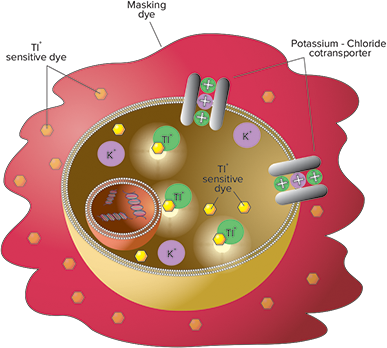
***Figure 1. The FLIPR Potassium Assay Kit principle.*Increase in cytosolic TI+is detected by FLIPR or FlexStation systems using thallium-sensitive dye indicators.
The FLIPR Potassium AssayExplorer Kit (Molecular Devices Cat. # R8222) contains a Tl+-sensitive dye, masking dye for homogenous operation, 200 mM K2SO4, 50 mM Tl2SO4, 5X chloride-free buffer, and HBSS with 20 mM HEPES buffer. The kit is sufficient for ten 96-, 384-, or 1536-well plates. The assay workflow is shown in Figure 2.
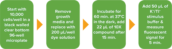
Figure 2. FLIPR Potassium Assay Kit workflow on the FLIPR Tetra High-Throughput Cellular Screening System.
Experimental procedures
Cell culture and transfection
HEK293T (ECACC Cat. # 12022001) cells were grown to 80–90% confluence in T75 flasks containing growth medium (DMEM supplemented with 10% FBS and 2 mM L-Glutamine). Cultures were maintained at 37°C in the presence of 5% CO2. Cells were passaged every 3–4 days at a ratio of 1:6. Two days before the assay, cells were transiently transfected with human KCC2 (hKCC2) using Lipofectamine 2000 (Thermo Fisher Cat. # 11668027) according to the manufacturer’s instructions. The transfection mixture was mixed with growth medium, plated directly into a 96-well, black-walled, clear bottom plate at 10,000 cells/well and incubated at 37°C and 5% CO2for 48 hours.
KCC2 inhibition assay
Growth medium was removed from the plates which were then incubated with either FLIPR PotassiumAssay Kit or BTCAM (2 µM) for one hour at 37°C in the dark. The loading buffer contained 10 µM bumetanide to reduce the endogenous NKCC Tl+influx/transport signal, and 0.1 mM ouabain, a Na+/K+-ATPase blocker, to inhibit ATP-dependent sodium-potassium exchange across the cell membrane. Following the dye loading step, cells incubated with BTC-AM were washed to remove excess dye. The KCC2 inhibitors were added and incubated for the last 45 minutes of the dye loading time (FLIPR Potassium Assay Kitonly). Optimized stimulus buffer (9 mM K+/ 0.9 mM Tl+) was added to the well during detection on the FLIPR®Tetra System using the 470–495 nm excitation LEDs and 515–575 nm emission filter. Data files were imported into SoftMax®Pro Data Acquisition and Analysis Software for subsequent analysis using the Import Feature (Figure 3), available for SoftMax Pro 6.4.1 or higher. The initial rate (Vmax in units per second) was calculated from the first 10 seconds of data post stimulus addition.

Figure 3. SoftMax Pro 6.4.2 import workflow.
Results
After loading hKCC2-transfected or mocktransfected HEK293 cells with the novel FLIPR Potassium Assay Kit or BTC-AM, we measured the increase in fluorescence triggered by the addition of external Tl+during detection on the FLIPR TetraSystem. The hKCC2-driven FLIPR signal exhibits a rapid increase over the first ten seconds after Tl+addition in cells loaded with the FLIPR Potassium AssayKit, followed by a slower increase and eventually a plateau phase. The increase was significantly higher (P < 0.001; Student’s t-test) in hKCC2- expressing cells, compared to mocktransfected cells as shown in Figure 4. The average Z factor was calculated as 0.60 ± 0.05 indicating a robust assay2. In contrast, with BTC-AM loaded cells the signal was much slower to develop, showed a large degree of variability and the magnitude of the signal was far lower. For these reasons, no further comparisons between the assay kit and BTC-AM were made.
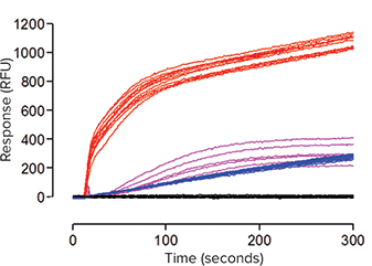
***Figure 4. Representative fluorescence signal traces (≥ 6 wells per treatment) obtained on the FLIPR Tetra System in HEK293 cells loaded with the FLIPR Potassium Assay Kit or BTC-AM.*Data show the effect of buffer only addition (–) or Tl+addition (–) in hKCC2 transfected cells, and Tl+addition (–) in mock transfected cells loaded with the FLIPR Potassium Assay Kit. For comparison, the effect of Tl+addition (–) in transfected cells loaded with BTC-AM is also shown.
A series of experiments were then carried out with hKCC2-expressing cells loaded with the FLIPR PotassiumAssay Kit to assess whether the Tl+influx assay could be used to detect modulators of hKCC2 activity. In each 96-well plate, cells were treated with either vehicle control or 30 µM R-(+)-DIOA (an alkanoic acid that has been shown to be a potent and selective inhibitor of K+/Cl-cotransporters). As seen in Figure 5, the resultant signals from the K+/Tl+-evoked increase in fluorescence are very consistent from well to well, and the separation between the control and R-(+)-DIOA signals is significant (P < 0.001; Student’s t-test).
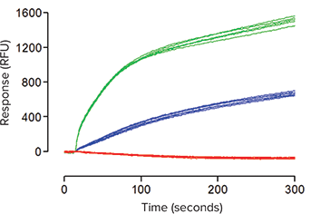
***Figure 5. Representative fluorescence traces obtained on the FLIPR Tetra System in hKCC2- transfected cells loaded with the FLIPR Potassium Assay Kit.*Data show the effect of buffer only addition (–) or Tl+addition in the absence (–) or presence (–) of 30 µM R-(+)-DIOA.
In the absence of inhibitor the K+/Tl+stimulus produced a Vmax of 53.9 ± 5.1 units per second, whereas in the presence of 30 µM R-(+)-DIOA this was reduced to 17.5 ± 1.9 units per second (Figure 6) where n ≥ 30 wells over four independent experiments.
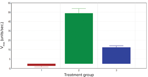
***Figure 6. The effect of different challenges on hKCC2-transfected cells.*Data shows the effect of three different treatment groups: 1. buffer only addition (■), and 2. Tl+/K+addition in the absence (■) or 3. presence (■) of 30 µM R-(+)-DIOA. Bars represent mean Vmax (units/sec.) ± SEM (n ≥ 30).
Conclusions
We have shown that the FLIPR PotassiumAssay Kit can be used to measure the functional activity of the hKCC2 cation-chloride cotransporter using a homogeneous, no-wash protocol. The assay kitdisplayed a large assay window and excellent reproducibility, and was able to successfully detect a known modulator of hKCC2 activity. Compared to traditional dyes such as BTC-AM, the simplified protocol and robust assay quality, in combination with the high-throughput capability of the FLIPR TetraSystem, provides a powerful solution to screen for modulators of the hKCC2 cotransporter.
Acknowledgements
Andrea Townsend-Nicholson and Stephanie Schorge, University College London, and Simon Lydford, Molecular Devices (UK) Ltd
References
- Kahle KT, Staley KJ, Nahed BV, Gamba G, Hebert SC, Lifton RP, et al. Roles of the cation-chloride cotransporters in neurological disease. *Nat Clin Pract Neurol.*2008;4:490-503
- Zhang JH, Chung TD & Oldenburg KR. A Simple Statistical Parameter for Use in Evaluation and Validation of High Throughput Screening Assays. *J Biomol Screen.*1999;4:67-73.
Learn more >>
介绍
氯化钾转运体成员 5 ( SLC 12 A 5 或 KCC 2 ) 是由 SLC 12 基因家族编码的九种阳离子氯 离子共转运体 (CCCs) 之一,是神经元中唯 一优先表达的 CCC 转运体。KCC 2 在中枢 神经系统的正常运作中起着至关重要的作 用,在维持神经元细胞内 Cl‒ 浓度 ([Cl‒ ] i ) 方 面起着关键作用。KCC 2 参与了许多神经 元过程的控制,其受损的活动已在脊髓损 伤后的癫痫、神经性疼痛和痉挛中得到证 实。这表明 KCC 2 活性或表达的阳性调节 剂可能为 KCC 2 功能缺陷引起的神经系统 疾病提供有效的治疗。
FLIPR 钾离子检测试剂盒含有一种新型的高 灵敏度铊 (Tl+ ) 指示剂染料,该染料通过钾 离子通道与 Tl+ 结合后产生明亮的荧光。 信号强度与细胞上钾离子通道开放数量成 正比;因此它可以作为钾离子通道活性的 替代指标。该试剂盒还采用了 Molecular Devices 专有的屏蔽染料,以减少背景荧 光,从而提高信号/背景比。由于 Tl+ 也由 阳离子-氯离子共转运体转运,因此 FLIPR 钾检测试剂盒可以通过测量 KCC 2 介导的 Tl+ 转运/流入的初始速率,轻松评估化合 物对 KCC 2 活性的影响。
实验原理
与非均质检测相比,该检测试剂盒的 Tl+ 指 示剂染料具有一个扩大的信号窗口。BTC - am 是一种 Ca2+ 指示剂,由于 Tl+ 离子也能 增强 BTC 的荧光,因此也被用于钾离子通 道的监测。在染料加载过程中,Tl+ 指示剂 染料以乙酰氧基甲基酯 (AM) 的形式通过细 胞膜被动扩散进入细胞。细胞质酯酶裂解 AM 酯,释放出活性的成氟形式。检测试剂 盒中含有一种专有的细胞外屏蔽染料,以 减少背景荧光 ( 图 1 )。为了激活氯化钾转 APPLICATION NOTE 运体,在存在或不存在测试化合物 ( 如 KCC 2 抑制剂 ) 的情况下,用 K+ 和 Tl+ 的混合物 刺激细胞。在使用的检测条件下,荧光信 号的增加代表了 Tl+ 通过共转运体特异性地 进入细胞,因此代表了对共转运体活性的 功能测量。共转运体活性的调节是通过包 含 KCC2 抑制剂来实现的。
***图 1 FLIPR 钾离子检测试剂盒原理。*细胞胞质 TI+ 的增加可通过 FLIPR 或 FlexStation 系统使用铊敏感染 料指示剂检测到。
FLIPR 钾离子检 测试剂盒 (Molecular Devices,Cat# R8222) 含有 Tl+ 敏感染料,用于均相操作的屏蔽染 料,200 mm K2 SO4 , 50 mm Tl2 SO4 , 5X 无氯 缓冲液,以及含有 20 mm HEPES 的 HBSS 缓冲液。该试剂盒可用于 10 个 96-、384 - 或 1536 孔板。检测工作流程如图 2 所示。
图 2 FLIPR 钾离子检测试剂盒在 FLIPRTetra 高通量实时荧光检测分析系统上的工作流程。
实验流程
细胞培养和转染
HEK 293T (ECACC Cat. # 12022001) 细胞 在含有生长培养基 ( 补充有 10% FBS 和 2 mm L - 谷氨酰胺的 DMEM ) 的 T75 烧瓶 中生长至 80 - 90% 汇合。在 5% CO2 的 存在下,培养物保持在 37℃。细胞每 3 - 4 天传代一次,传代率为 1 : 6。实验前两天用 Lipofectamine 2000 (Thermo Fisher Cat. # 11668027) 转染人 KCC 2 (hKCC 2) 细胞。 根据制造商的说明。转染混合物与生长培 养基混合,直接在 10000 个细胞/孔的 96 孔黑壁透明底板上铺板培养,37℃、5% CO2 孵育 48 小时。
KCC 2 抑制实验
孔板中的生长培养基被移除,然后添加 FLIPR 钾离子检测试剂盒或 BTCAM (2 μM) 在 37°C 条件下避光孵育一小时。加载的缓 冲液中包含 10 μM 布美他尼,用于减少内 源性 NKCC Tl+ 流入/传输信号,同时含有 0.1 mM 乌本苷,作为一种 Na+ /K+ - ATP 酶 阻断剂,用于抑制 ATP 依赖的细胞膜钠钾 交换。在加载染料之后,清洗 BTC - AM 共 孵 育 的 细 胞 以 去 除 多 余 的 染 料 。 添 加 KCC 2 抑制剂,并在染料加载时间的最后 45 分钟孵育 ( FLIPR 钾离子检测试剂盒处 理组 )。在 FLIPRTETRA 系统上检测时, 在孔中加入优化的刺激缓冲液 (9 mm K+ /0.9 mM Tl+ )。Tetra 系统采用 470 - 495 nm 激 发 LED 和 515 - 575 nm 发射滤光片。数据 文件被导入 SoftMaxPro 数据采集和分析 软件中,以便使用导入特性 ( 图 3 ) 进行后 续分析,该特性可用于 SoftMaxPro 6.4.1 或更高版本。初始速率 ( Vmax,单位为每 秒 ) 是根 据刺激后的前 10 秒数据计算出来 的。
图 3 SoftMax Pro 6.4.2 导入流程。
结果
用新型 FLIPR 钾离子试剂盒或 BTC - AM 加 载 hKCC 2 转染或模拟转染的 HEK 293 细胞 后,我们测量了在 FLIPR Tetra 系统上检 测时添加外部 Tl+ 引起的荧光增加。在装有 FLIPR 钾离子检测试剂盒的细胞中加入 Tl+ 后,hKCC 2 驱动的 FLIPR 信号在最初 10 秒内呈现快速增长,随后增长较慢,最终 进入平稳期。与模拟转染细胞相比, hKCC 2 转染细胞荧光信号显著更高 (P < 0.001;Student’s t-test),如图 4 所 示。平均 Z 因子计算为 0.60 ± 0.05,表明 实验结果的稳定和可靠 2。与此相反,BTC - AM 加载细胞的信号增加较慢,表现出很大 的变异性,信号的大小要低得多。由于这 些原因,没有进一步比较分析试剂盒和 BTC - AM。
***图 4 FLIPR Tetra 系统上获取的代表性荧光信号轨迹 ( 每个处理组 ≥ 6 孔 ), HEK 293 细胞加载了 FLIPR 钾离子检测试剂盒或 BTC - AM。*数据显示,在 hK CC 2 转染细胞中,仅添加缓冲液 (-) 或添加 Tl+ (-),以及在加载 FLIPR 钾离子检测试剂盒的模拟转染细胞中,仅添加缓冲液 (-) 或添加 Tl+ (-) 的效 果。同时,还比较了添加 Tl+ (-) 对转染细胞中加载 BTC - AM 的影响。
然后我们用加载有 FLIPR 钾离子试剂盒的 表达 hKCC 2 的细胞进行了一系列实验,以 评估 Tl+ 内流试验是否可以用于检测 hKCC 2 活性的调节剂。在每一个 96 孔板中,细 胞被分为对照溶液组或 30 μM R - (+) - DIOA ( 一种烷酸,已被证明是 K+/Cl - 协同转运 体的一种有效的选择性抑制剂 ) 组。从图 5 中可以看出,K+/Tl+ - 诱发的荧光增强所 产生的信号在孔与孔之间非常一致,对照 组信号与 R - (+) - DIOA 信号显著区分 (P < 0.001; Student’s t - test)。
***图 5 FLIPR Tetra 系统上获取的代表性荧光信号轨迹 ,hK CC2 - 转染细胞加载了 FLIPR 钾离子检 测试剂盒。*数据显示了在 30 μM R - (+) - DIOA 存在或不存在情况下,只添加缓冲液 (‒) 或添加 Tl+ 的 效应。
在没有抑制剂的条件下, K+/Tl+ 刺激产生 Vmax 53.9 ± 5.1 (units/second),而 在 30 μM R - (+) - DIOA 条件下,超过四次独立实 验中总计 n ≥ 30 孔的结果表明 (Figure 6 ), Vmax 减少至 17.5 ± 1.9 (units/second)。
***图 6 不同条件对 hkcc 2 转染细胞的影响。*数据显示了三种不同处理组的结果:1.只添加缓冲溶液 (n), 和 2. 在没有 (n) 或 3. 存在 (n) 30 μM R - (+) -DIOA 的情况下添加 Tl+ /K+。柱状图代表的意思是Vmax (units/sec.) ± SEM (n ≥30)。
结论
我们已经证明 FLIPR 钾离子检测试剂盒可 以 使 用 均 匀 的 、 不 清 洗 的 方 案 来 测 量 hKCC 2 阳离子 - 氯离子共转运体的功能活 性。该检测试剂盒显示出较大的检测窗 口 和良好的重现性,能够成功地检测到 已知的 hKCC 2 活性调节剂。与传统染料如 BTC - AM 相比,简化的实验方案和稳健的 检测质量,结合 FLIPR Tetra 系统的高通 量能力,为筛选 hKCC 2 共转运体调节剂提 供了一个强大的解决方案。
致谢
Andrea Townsend-Nicholson and Stephanie Schorge, University College London, and Simon Lydford, Molecular Devices (UK) Ltd.
参考文献
- Kahle KT, Staley KJ, Nahed BV, Gamba G, Hebert SC, Lifton RP, et al. Roles of the cation-chloride cotransporters in neurological disease. *Nat Clin Pract Neurol.*2008;4:490-503
- Zhang JH, Chung TD & Oldenburg KR. A Simple Statistical Parameter for Use in Evaluation and Validation of High Throughput Screening Assays. *J Biomol Screen.*1999;4:67-73.
Learn more >>