Superamento delle difficoltà associate all’imaging 3D ad alto rendimento
Grazie ai recenti progressi nelle tecnologie di imaging, siamo attualmente in grado di osservare e analizzare complesse reti cellulari in tre dimensioni. Attraverso l'imaging 3D, siamo in grado di acquisire e analizzare le interazioni in cellule e tessuti con maggiore dettaglio e maggiore precisione. Tuttavia, l’imaging 3D è un processo complicato, implicante molti livelli di complessità, dai lunghi tempi di scansione delle immagini alla bassa risoluzione e agli strumenti di analisi inadeguati.
Ecco un riepilogo delle difficoltà comuni affrontate nell’imaging cellulare 3D e di come superarle con l’aiuto dei prodotti di Dispositivi Molecolari.
Luce fuori fuoco
Ottenere immagini 3D nitide può essere difficile, specialmente se si utilizza la microscopia a campo largo. Nonostante forniscano una rapida acquisizione delle immagini, la microscopia in campo largo non riesce a eliminare i segnali fuori fuoco. Questo ti lascia con immagini sfocate con luce intensa fuori fuoco, ostacolando l'analisi delle immagini.
L'alternativa è un micrometro confocale con un foro che riduce la sensibilità alla luce fuori fuoco quando ci si allontana dall'area di messa a fuoco, generando immagini più nitide. Il problema si verifica quando un singolo foro deve eseguire la scansione dell'intera immagine, il che richiede tempo.
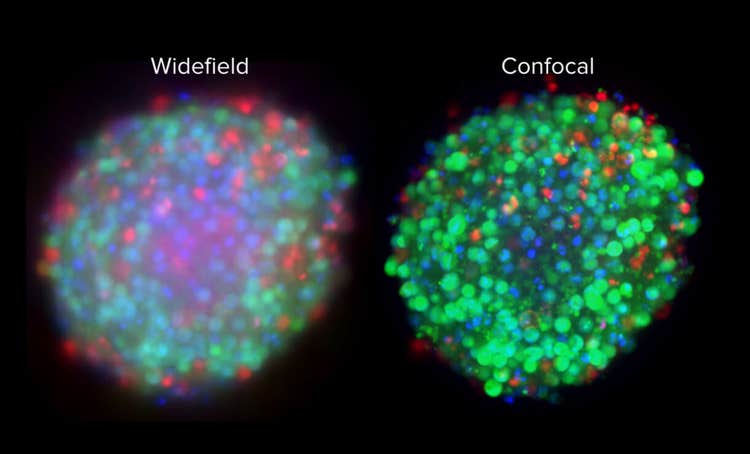
È qui che un microscopo confocale a disco rotante (SCDM) può fungere da aggiornamento. Invece di un singolo foro, un SDCM consiste di centinaia di fori che ruotano rapidamente per eseguire la scansione dell'immagine, portando a una migliore risoluzione in breve tempo. La tecnologia confocale del disco di rotazione AgileOptix ne è un esempio perfetto.
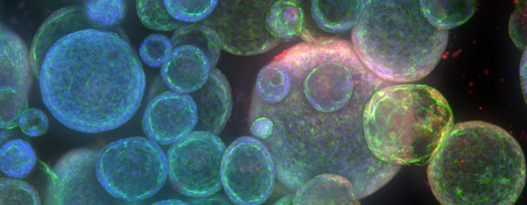
Il sistema di diagnostica per immagini ad alto contenuto microconfocale ImageXpress® è dotato della tecnologia confocale del disco di rotazione, una tecnologia che fa un ulteriore passo avanti con una sorgente luminosa laser ad alta intensità per una maggiore penetrazione dei tessuti, in modo da poter ottenere immagini ad alta risoluzione anche da campioni di tessuto spessi. Di conseguenza, è possibile ottenere un'immagine più nitida con una migliore visibilità cellulare e un aumento di almeno il 30% della conta cellulare.
Lungo tempo di acquisizione
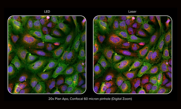
Prima delle tecnologie di scansione laser 3D, l'acquisizione di immagini di alta qualità richiedeva esposizione prolungata, il che era una difficoltà a causa delle immagini 3D che si allontanavano dal centro del pozzo. Oggi, i microscopi confocali affrontano questo problema grazie all'implementazione di sorgenti laser ad alta intensità.
Le sorgenti ad alta intensità hanno diversi vantaggi che non solo migliorano la qualità dell'immagine, ma aumentano anche la velocità di scansione riducendo il tempo di esposizione.
Ad esempio, il sistema di imaging ad alto contenuto ImageXpress® Confocal HT.ai è dotato di una sorgente luminosa laser a sette canali con otto canali di imaging in grado di ridurre il tempo di esposizione fino al 75%, il che aumenta la velocità di scansione di due volte in generale.
Questo è particolarmente utile con l’imaging delle proteine fluorescenti gialle o ciane che richiede l’imaging di reti proteiche complesse con eterogeneità intercellulari e intracellulari.
Messa a fuoco automatica affidabile
Mantenere la concentrazione sui campioni con fluttuazioni termica e meccanica è impegnativo e può interrompere in particolare l'imaging time-lapse. Ecco perché il tuo micrometro dovrebbe rilevare e stabilizzare automaticamente i piani focali. I sistemi di autofocus basati su fotocamera digitale richiedono molto tempo perché esiste un processo di pre-messa a fuoco con una ricerca intricata su una vasta gamma di punti di messa a fuoco.
Con i recenti avanzamenti nelle tecnologie di autofocus laser, è possibile accelerare la messa a fuoco automatica su vari tipi di piastre, tra cui organo su chip e fondo a U.
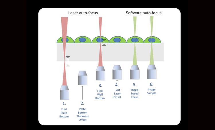
In particolare, un sistema di automessa a fuoco ibrido che comprende l'automessa a laser e l'immagine genererà i migliori risultati. Questo tipo di autofocus è molto più veloce perché il laser viene lampeggiato solo una volta per pozzetto. Tuttavia, è ancora possibile ottenere benefici dalla compatibilità dei campioni della messa a fuoco basata su immagini. Ancora più importante, la quantità minima di esposizione laser significa che si sta riducendo al minimo il rischio di fototossicità, che è fondamentale nei test su cellule vive.
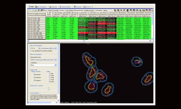
Collo di bottiglia dell'analisi
I set di dati sono spesso grandi e complessi, quindi il completamento dell'analisi può richiedere ore. Superare questo ostacolo richiede strumenti di analisi delle immagini all'avanguardia con elaborazione accelerata, classificazione accurata delle immagini e una semplice interfaccia utente.
Gli strumenti di analisi delle immagini di Molecular Devices si distinguono per le loro caratteristiche che risolvono una serie di esigenze di analisi.
Ad esempio, sapevi che potresti accelerare l'analisi time-lapse fino a 40 volte con l'elaborazione parallela multi-thread? Questo è diventato una realtà con il nostro software di acquisizione e analisi delle immagini ad alto contenuto MetaXpress® per l’imaging 2D e 3D. Un'altra difficoltà nell'analisi delle immagini è la classificazione delle popolazioni di cellule per una Caratterizzazione cellulare completa. Ecco dove entra in gioco il macchina learning. Il nostro software di analisi delle immagini IN Cartaý consente la classificazione automatica della fenotipizzazione in profili cellulari grandi e complessi.
In entrambi gli strumenti di analisi, è possibile eseguire processi con una varietà di campioni in pochi minuti con un'esperienza utente eccellente.
Difficile penetrazione della luce
Forse una delle attività più difficili nell’imaging 3D è la penetrazione dei tessuti, che è fondamentale per ottenere informazioni sul comportamento biologico complesso del campione. Se il micrometro ha una profondità di penetrazione limitata, la qualità dell'immagine diminuirà man mano che la luce viene sparsa o assorbita in campioni di tessuto spessi.
Come accennato in precedenza, la microscopia confocale può fornire una migliore risoluzione, ma ulteriori sviluppi possono tradurre questo successo in una profonda penetrazione dei tessuti.
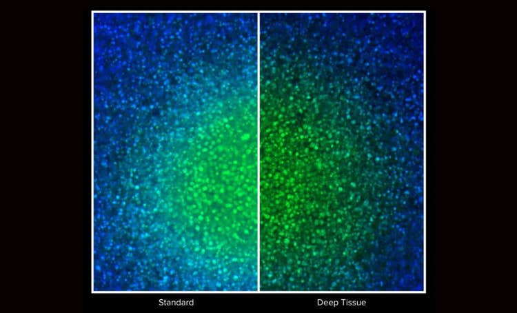
La capacità di contrasto del microscopio è fondamentale per la soppressione della luce fuori fuoco, il che consente una migliore rilevazione della fluorescenza emessa dal campione. Ciò può essere ottenuto mediante un foro che rifiuta il segnale fuori fuoco. Il contrasto può essere raggiunto ancora più velocemente se si dispone di più fori che sono in comunicazione costante tra loro durante la scansione delle immagini.
Infine, la combinazione di microscopia confocale con ottica laser ad alta intensità consente una maggiore penetrazione inviando lunghezze d’onda più lunghe di luce per eccitare i campioni di fluorescenza senza danneggiare il campione o diffondere la luce.
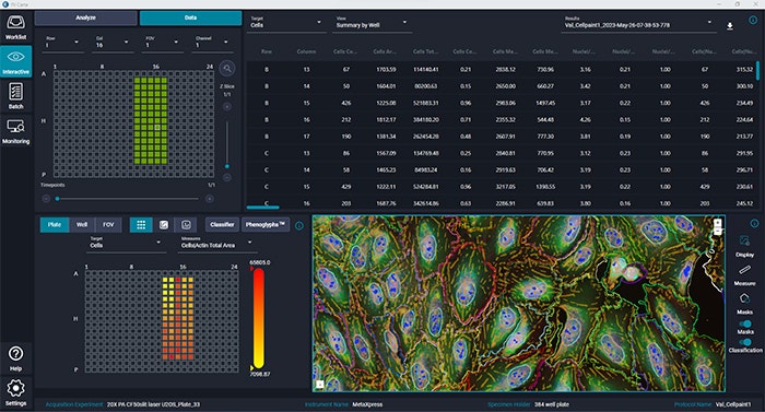
Data-mining ad alto contenuto
Immaginiamo di provare a visualizzare i dati multi-pozzetto o multi-piastra. Con un software di imaging 3D autonomo, probabilmente è necessario passare da un'applicazione all'altra per visualizzare più anteprime di immagini e mappe di calore.
Per rendere più pratico l'estrazione dei dati, è necessario disporre di un software informativo in grado di integrare l'acquisizione delle immagini con l'analisi delle immagini e l'informatica in modo da poter analizzare i dati direttamente dall'immagine originale. Con questo scopo in mente, i dispositivi Molecolari hanno sviluppato il software informativo ad alto contenuto Acuity .
Il software è dotato di una funzione di zoom interattivo che consente di navigare senza problemi tra immagini e dati numerici. Con questa tecnologia, si è al posto del conducente quando si determina il livello di dettaglio, che si tratti di zoomare in un'immagine singola di alta qualità o di una visualizzazione complessiva di tutte le immagini e i dati su un unico schermo.
Oltre alla navigazione all’ultimo grido, il software AcuityXpress offre una pletora di strumenti di analisi e calcolo per interpretare dati complessi e più parametri contemporaneamente.
Conclusione
L’imaging 3D può portare informazioni più profonde sul comportamento complesso dei tuoi campioni; tuttavia, comporta alcune difficoltà. In Molecular Devices, ci adoperiamo per accompagnarvi in ogni fase del processo di diagnostica per immagini per guidarvi verso la vostra soluzione specifica: dall’acquisizione di immagini più nitide in pochi minuti all’organizzazione di queste immagini in un sistema software centralizzato, vi aiuteremo a far avanzare la ricerca.
per saperne di più su High-Content Screening con tecnologia AgileOptix e la sua combinazione di potenti laser a stato solido, obiettivi di immersione in acqua, sensore scientifico CMOS e doppio disco rotante con cinque diverse geometrie di disco. Oppure contattaci per ulteriori informazioni.