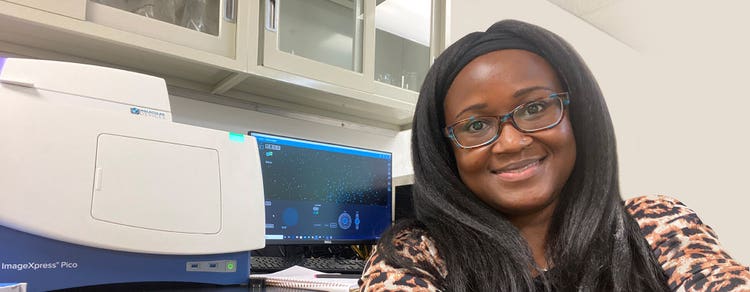
Vi presentiamo il nostro field applications scientist: Cheryl Bell
Cheryl Bell parla di come effettuare la transizione dalla micropsia manuale alla micropsia automatica

Cheryl, ci dica del suo background e di come è arrivato/a a Molecular Devices.
Ho completato i miei studi universitari presso la Jackson State University di Mississippi, dove mi sono conferito in biologia pre-med. Ho fatto una lezione di genetica come parte del programma e l’ho amata assolutamente! Questo mi ha portato a perseguire il mio dottorato in genetica e genomica presso l’Università del Connecticut, dove mi sono concentrato sulla terapia di prevenzione dell’HIV per i maschi non circoncisi. Il progetto è stato collegato a una sperimentazione clinica che ha richiesto anni di valutazione microscopica dei campioni per validare l'efficacia del farmaco terapeutico che stavamo studiando. Da lì, ho completato la mia formazione post-dottorato in biologia cellulare presso l’Università di Pittsburgh, dove ho studiato le proteine di giunzione spaziale con l’obiettivo di applicare i miei insegnamenti al campo della guarigione delle lesioni.
Durante il corso dei miei studi, ho utilizzato diversi tipi di tecniche di microscopia, tra cui la microscopia elettronica a fluorescenza, confocale e a trasmissione. Il mio obiettivo in quel momento era quello di diventare un docente e di eseguire il mio laboratorio. Verso la fine del mio corso di formazione post-dottorato, i miei piani hanno fatto una svolta quando uno dei miei amici della scuola di istruzione mi ha contattato per una posizione aperta per uno scienziato di applicazioni di campo di imaging cellulare presso Molecular Devices. Ho finito per ottenere la posizione ad aprile 2018.
Effettuare la transizione dal mondo accademico a un ambiente aziendale mi ha consentito di trasferire le conoscenze e le competenze che ho acquisito dai miei anni di ricerca per aiutare i clienti a rispondere alle loro domande scientifiche attraverso la micropsia.
Ci puoi dire di più sul tuo ruolo in dispositivi Molecolari?
Attualmente ho sede a Chicago, Illinois. Nel mio ruolo di Field Applications Scientist, fornisco supporto tecnico e di vendita per il nostro sistema di imaging cellulare automatico ImageXpress® Pico . Ciò include la conduzione di sessioni didattiche o sessioni individuali con i clienti, in cui rispondo a domande tecniche relative al prodotto o ad applicazioni specifiche.
Il sistema ImageXpress Pico è stato lanciato circa nello stesso periodo in cui sono entrato a far parte di Molecular Devices. Da allora la piattaforma si è evoluta abbastanza, con miglioramenti come il controllo ambientale ,il confocale digitale e l'anteprima dal vivo .
La maggior parte dei clienti del sistema ImageXpress Pico sono utenti principianti o avanzati di microscopie?
Direi che metà dei nostri clienti sono utenti principianti che erano abituati a utilizzare la microscopia manuale prima di passare al sistema ImageXpress Pico . Questi clienti stavano cercando di aumentare la rendimento e ridurre la quantità di tempo impiegata per l'acquisizione di immagini e l'analisi dei dati. Utilizzando questo sistema, questi clienti sono ora in grado di ridurre la parte di imaging del loro carico di lavoro da ore a minuti. Sono anche in grado di generare più dati dal loro esperimento.
Le funzioni classiche che si dovrebbero eseguire su un microscopio manuale sono semplificate con l'imaging automatico. In questo modo si elimina la necessità di apportare modifiche durante l'acquisizione dell'immagine. Fondamentalmente, il sistema ImageXpress Pico aiuta i clienti a ottimizzare il flusso di lavoro di diagnostica per immagini e analisi cellulare.
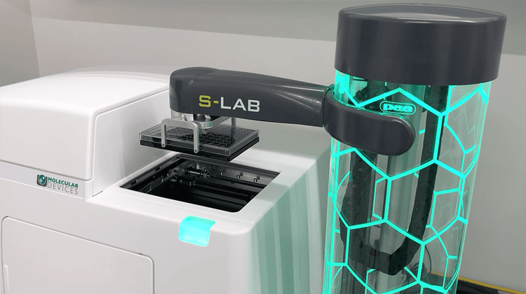
Considererei la parte rimanente dei nostri clienti come utenti avanzati che hanno già familiarità con l'imaging automatico. Questi individui si rivolgono al sistema ImageXpress Pico per la sua qualità dell’immagine facile da usare e ad alta risoluzione, nonché per la sua versatilità nell’accogliere diversi tipi di campione e articoli da laboratorio. Gli utenti più avanzati di solito sfruttano le nostre soluzioni di personalizzazione e automazione. Ciò può comportare l’aggiornamento a funzioni aggiuntive nel software CellReporter smk o la combinazione con altri software di dispositivi Molecolari come il software di acquisizione e analisi delle immagini ad alto contenuto Meta smk o il software SoftMax . All'interno di queste piattaforme software, possono attivare determinate funzioni come l'acquisizione di Z-stack o Digital Confocal . Inoltre, possono utilizzare le nostre ultime personalizzazioni con un braccio robotizzato e un’incubatrice.
In pratica, il sistema ImageXpress Pico è progettato per accogliere una vasta gamma di utenti. Poiché lo strumento è così facile da usare, è ideale per le strutture con un elevato tasso di ricambio, consentendo al nuovo personale di mettersi in funzione molto rapidamente.
Quali sono alcune delle difficoltà comuni che si osservano con i clienti in relazione all'imaging automatico?
Ritengo che una difficoltà comune per i nostri clienti sia la gestione dei dati. L'imaging automatico genera molti dati. Questo può essere sulla scala di gigabytes o persino terabytes. In molti casi, dobbiamo guidare i clienti su come salvare i dati in diversi formati compatibili con i loro sistemi.
Un'altra difficoltà per i clienti è determinare i tipi di piastre o macchie che sono ottimali per la loro particolare applicazione o test. Ad esempio, il modo in cui i clienti sono abituati a preparare i loro campioni potrebbe non essere ideale per l'imaging automatico. Lavoro con i clienti per apportare le regolazioni necessarie nella preparazione dei campioni in modo che possano facilmente rendere automatico il processo di diagnostica per immagini e generare i dati di cui hanno bisogno.
Ho anche osservato che i clienti stanno diventando sempre più interessati all'imaging di modelli di celle 3D. In questi casi, li aiuteremo a identificare il sistema di diagnostica per immagini che meglio soddisfa le loro esigenze di ricerca. Il nostro portafoglio di prodotti per l’imaging è configurato in modo da facilitare la transizione dei clienti dal sistema ImageXpress Pico a un modello più alto, come il nostro ultimo sistema di imaging ad alto contenuto ImageXpress® Confocal HT.ai, mentre la loro ricerca continua a migliorare.
Ci sono casi in cui i clienti possono scegliere di utilizzare il sistema ImageXpress Pico insieme al sistema ImageXpress Confocal HT.ai come parte del loro flusso di lavoro?
Sì, assolutamente. Abbiamo clienti che utilizzano il sistema ImageXpress Pico per eseguire test di convalida o immagini più semplici, dove devono eseguire piastre 96a pozzetto ad alta rendimento. Quando i clienti devono acquisire immagini ad alta risoluzione per un’applicazione specifica, possono eseguire la piastra nel sistema ImageXpress Confocal HT.ai . La facilità di utilizzo del sistema ImageXpress Pico aiuta i clienti a superare i loro esperimenti più rapidamente e infine ad acquisire i dati necessari.
Abbiamo anche clienti che utilizzano i nostri lettore di micropiastre insieme al sistema ImageXpress Pico per alcune applicazioni. È possibile trasferire facilmente i dati acquisiti dal sistema ImageXpress Pico al software SoftMax Pro che viene utilizzato con i nostri lettore di micropiastre. Ritengo che il sistema ImageXpress Pico sia la piattaforma intermedia che aiuta i nostri clienti ad arrivare dove devono andare in modo più efficiente.
Ci sono applicazioni o test particolari che ti entusiasmo davvero per quanto riguarda l'imaging?
Quello che mi piace di più del mio ruolo di scienziato delle applicazioni è che posso vedere i nostri sistemi utilizzati per una vasta gamma di applicazioni. La capacità di acquisire immagini di organismi come vermi, fitoplancton, rotifer e vedere la biologia molecolare al suo centro in tempo reale è molto entusiasmante per me! Sono anche emozionato di lavorare con campioni come il cervello di topo e il pesci zebra.
Sto vedendo i clienti sviluppare alcuni test all'avanguardia attraverso la loro personalizzazione degli articoli da laboratorio. Possono creare tutti i tipi di articoli da laboratorio progettati per coltivare determinati tipi di cellule o consentire ai batteri di coltivare le collettività. Ad esempio, ho clienti che stanno creando canali multipli per la crescita dei neuroni. Questo crea un ambiente che è più rappresentativo del modo in cui i neuroni operano in vivo . Dal momento che queste piattaforme sono più rilevanti dal punto di vista biologico, consentono ai ricercatori di sondare ulteriormente i loro campioni e di disegnare dei farmaci migliori per diverse condizioni come l’Alzamper. Ho un altro cliente che sta studiando le comunità batteriche nell’intestino in modo che possano ottenere maggiori informazioni su come disegnare probiotici per gli individui che sono affetti da problemi gastrointestinali. I modi innovativi in cui i clienti stanno creando scenari biologici reali per migliorare il modo in cui comprendiamo la salute e le malattie mi soffia davvero la mente!
I sistemi di imaging sono compatibili con gli articoli da laboratorio personalizzati che i clienti stanno creando?
I nostri sistemi di imaging sono compatibili con una vasta gamma di articoli da laboratorio. Quando necessario, il nostro team collaborerà con i clienti per configurare le nostre piattaforme hardware e/o software per soddisfare i loro requisiti di laboratorio unici.
https://share.vidyard.com/watch/261PZTTCKAUFtD7skdrw2M
Quali avanzamenti futuri prevedete per la micropsia automatica?
La microscopia automatica aprirà molte vie per diverse aree di ricerca. Ad esempio, se ci si trova nel campo dell’organoide, si stanno coltivando questi campioni nel tempo. È necessario acquisire ripetutamente immagini dei campioni in momenti diversi durante il processo di crescita. Di recente ho acquisito un campione che aveva 160 giorni. Mentre immaginiamo campioni che stanno aumentando di dimensioni, avremo bisogno di piattaforme che possano vedere attraverso questi tipi di strutture. Prevedo che dovremo sviluppare una tecnologia che sia un integratore tra la microscopia e la scansione CAT. Questo ci consentirebbe di esaminare ancora più in profondità il campione.
Lungo queste linee, ritengo che il sistema ImageXpress Pico continuerà a migliorare e vedremo miglioramenti che aiutano a migliorare ulteriormente lo sviluppo di test e a semplificare il flusso di lavoro di diagnostica per immagini.
Guardando indietro alla tua carriera, c'era qualcosa in particolare che ha suscitato il tuo interesse per la tecnologia?
Dall’infanzia ero interessato al funzionamento interno del corpo umano, in particolare per quanto riguarda gli stati normali e malati. Ritengo che questa curiosità sia nata da mia sorella, che ha sviluppato una condizione autoimmune da teenager. Un giorno rifletterei su cosa potrebbe causare la salute di una persona come mia sorella e poi mi ammalerei il giorno successivo. Questo ha davvero suscitato il mio interesse per la tecnologia.
Quali sono alcuni dei tuoi interessi al di fuori del lavoro?
Mi piace trascorrere del tempo con i miei tre figli. Ora che sono più vecchi, trovo di avere più tempo per prendere nuovi hobby. Mi considererei un ambiverso. Quando sono fuori con gli amici o lavoro con i colleghi, sono molto estroverso. Quando sono a casa, mi piace trascorrere un po’ di tempo da solo a leggere o ascoltare la musica. Una delle mie attività di tutti i giorni preferite è la corsa. Ritengo che sia una forma di meditazione per me e sono fiero di aver completato la mia prima maratona l'anno precedente.

Per per saperne di più sulle nostre piattaforme di imaging automatico, consultare la nostra pagina Sistemi di imaging cellulare.